Cancer research and Leica Laser microdissection!

Cancer research and Leica Laser microdissection!

Anatomy - Pathology / Molecular Diagnostics / Cell Biology

December 18, 2018

DNA mutations lead to abnormal or missing functional proteins, which can cause cells to multiply uncontrollably and become cancerous. The extraction of pure tumor material is extremely important to understand the underlying mutation for a specific cancer type.

Leica Laser Microdissection systems allows the extraction of purely cancerous tissue without contaminating your sample with healthy cells and improve the workflow by precisely cutting only the cells you are interested in.

  • Every step is done under visual control and proceeds without contamination with surrounding tissue.

  • A New LMD software 8.2 with improved usability and larger application range.

  • A powerful and easy-to-use software that contains all needed features for a successful laser microdissection process enabling the user to easily select, dissect and visualize dissectates.
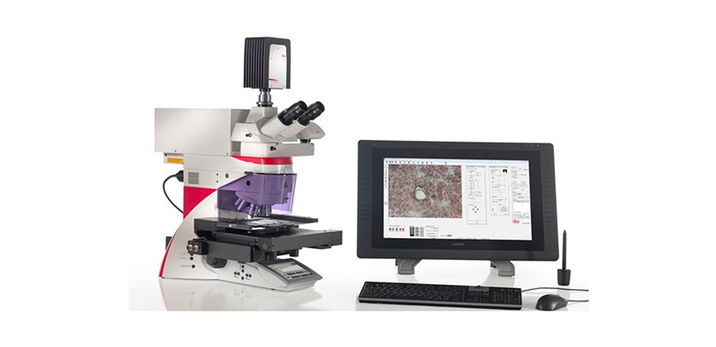
New Leica EC4 Microscope Camera

New Leica EC4 Microscope Camera

Anatomy - Pathology / Molecular Diagnostics / Cell...

Almost 10 years ago the Leica EC3 camera was introduced and today, Leica is pleased to announce the launch of the new Leica EC4 camera, the entry camera for microscope imaging with a c-mount interface.

December 10, 2015 More +
Antibody products against PEG from Abcam!

Antibody products against PEG from Abcam!

Anatomy - Pathology / Molecular Diagnostics / Cell...

Polyethylene glycol (PEG) is a synthetic polymer composed of repeating ethylene glycol (-CH2-CH2-O-) units. It has low toxicity, is strongly hydrophilic and has a large exclusion volume in aqueous solution. The covalent attachment of PEG (PEGylation) is commonly used to modify a variety of proteins and drugs. PEGylation can offer several benefits including decreased immunogenicity, increased bioavailability, and optimized pharmacokinetics5.


April 15, 2019 More +
Ready to make an enquiry Call us on: + 961 1 39 66 77
Or email us